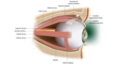
Human Eye Definition
The human eye is a complicated organ that permits us to see by detecting and focusing light. It's miles made up of numerous parts, which include the cornea, iris, student, lens, retina, and optic nerve. The cornea and lens paint together to focus light onto the retina, where its miles are converted into electrical alerts which can be sent to the brain via the optic nerve. The iris controls the quantity of light that enters the eye using adjusting the dimensions of the scholar. The eye additionally contains diverse structures that help to defend it and hold it lubricated, including the eyelids and tears. Typical attention is a completely complex organ that lets us peer.

Anatomy of the human eye
The human eye is a complex organ that is answerable for imagination and prescient. It's far divided into several elements, together with the cornea, iris, student, lens, retina, and optic nerve. The cornea is the clear outer floor of the eye, which helps to consciousness mild. The iris is the colored part of the eye that controls the size of the pupil, which regulates the amount of mild that enters the attention. The lens is located at the back of the iris and enables to similarly attention mild onto the retina. The retina is the innermost layer of attention and contains hundreds of thousands of mild-sensitive cells called rods and cones. Those cells convert the light that enters the eye into electric signals which might be sent to the brain via the optic nerve. The mind then translates these indicators as photographs.

Seen part of Eye
The visible part of the eye that may be seen from the outdoors the body is the sclera (white of the attention), the cornea, the iris (colored ring across the student), and the student. The eyelids and eyelashes additionally surround the attention and play a role in protecting it.

The floor of the eye

Front of the eye
The front of the attention refers back to the anterior part of the attention, which incorporates the cornea, iris, pupil, and lens. The cornea is a clean outer overlaying that facilitates to protect the attention and enables it to refract mildly. The iris is the colored part of the attention that surrounds the student and controls the amount of light that enters the attention. The scholar is the black circular opening within the center of the iris that lets in light to go into the attention. The lens is a clear structure positioned behind the iris that further allows consciousness light onto the retina.

The lower back of the eye
Retina shape and features
The again of attention, additionally known as the retina, is the light-sensitive layer of tissue that strains the interior of the attention. It consists of specialized cells referred to as rods and cones that detect light and send visible indicators to the mind thru the optic nerve. The retina is chargeable for converting light into electrical indicators that the brain can interpret as images. The macula, a small area placed near the center of the retina, is liable for presenting sharp, distinctive imaginative, and prescient. The retina additionally includes blood vessels that deliver oxygen and nutrients to the retina. Harm to the retina can purpose imaginative and prescient loss or blindness.

Inner a part of the eye
Internal Eye structure

Eye structure And feature
Attention is a complicated organ this is responsible for sensing mild and changing them into electrical indicators which can be despatched to the brain. The principal elements of attention include the cornea, iris, pupil, lens, retina, and optic nerve.
The cornea is the clear front surface of the eye that enables it to focus light. The iris is the colored part of the eye that surrounds the pupil, which controls the amount of mild that enters the attention. The lens is positioned behind the iris and facilitates similar focus mild onto the retina.
The retina is the innermost layer of the eye that includes thousands and thousands of mild-touchy cells known as rods and cones. These cells convert mild into electric alerts which can be despatched to the mind through the optic nerve.
In addition to those major parts, the attention additionally includes several different systems that assist to keep its fitness and function, inclusive of the vitreous humor, aqueous humor, and eyelids. The eyelids guard the attention against dirt and other overseas gadgets, even as the humor fluids offer nourishment and help to maintain the form of the eye.
Standard, attention is an incredibly complicated and precise organ that permits us to see and interpret the arena around us.


0 Comments